Blood Vessels Labeled Brain / The complex yet functional physiology of the circulatory ... / Label the blood vessels in the inferior view of the brain using the hints provided.
Blood Vessels Labeled Brain / The complex yet functional physiology of the circulatory ... / Label the blood vessels in the inferior view of the brain using the hints provided.. The blood vessels are the components of the circulatory system that transport blood throughout the human body. Label the blood vessels in the inferior view of the brain using the hints provided. Posterior communicating a internal carotid а. All cell types in the blood vessel wall can affect vessel diameter. Blood vessels innervate all tissues in vertebrates, enabling their survival by providing the necessary nutrients, oxygen, and hormonal signals.
• identification of blood vessels as arteries, capillaries or veins from the structure of their walls. Microscopically, it is formed by the endothelium of the blood vessel. Blood vessel endothelium is continuous with the inner tissue lining of organs such as the brain, lungs, skin, and heart. Label the blood vessels of the male pelvis using the hints provided. The brain and its surrounding blood vessels exist in a close relationship.
Master blood vessels with quizzes and diagrams | Kenhub from thumbor.kenhub.com The difference in the structural characteristics of arteries, capillaries and veins is attributable to their respective functions. Label the veins of the anterior forearm and hand. The precise relation between blood vessels and brain regions, reflecting the physiology and pathology of brain function directly and accurately, has remained largely unknown. The carotid arteries and the vertebral arteries anterior cerebral artery (aca): In the cerebral medulla, the arteries and veins of the right side of the body are controlled from the left side of the brain; Fill in the blanks with the appropriate words to describe blood flow from the heart. Label the blood vessels in the inferior view of the brain using the hints provided. They also take waste and carbon dioxide away from the tissues.
Blood is supplied to the brain through 2 major pairs of arteries.
Blood vessels in red in close communication with proliferating neuronal cells in the mouse cortex at embryonic day 10. Label the blood vessels in the inferior view of the brain using the hints provided. About 2 years ago updated: This is particularly important structure due to its clinical implications, which are discussed in more detail in the article. Blood vessels are critical to deliver oxygen and nutrients to all of the tissues and organs throughout the body. The brain and its surrounding blood vessels exist in a close relationship. Cerebral arterial circle anterior communicating posterior cerebral a middle cerebral al reset zoom. Fill in the blanks with the appropriate words to describe blood flow from the heart. Label the veins of the anterior forearm and hand. Towards the anterior side of the brain, those arteries are the internal carotid arteries. Blood travels from the heart in arteries, which branch into smaller and smaller vessels, eventually becoming arterioles. The two cell types ensure the integrity of the neural vasculature by maintaining the blood. The 500 ms patients, both adults and children, also underwent mri scans of the brain to measure iron deposits in surrounding areas of the brain.
Function and homeostasis of the brain relies on communication between its complex network of cells. Blood travels from the heart in arteries, which branch into smaller and smaller vessels, eventually becoming arterioles. The brain and its surrounding blood vessels exist in a close relationship. He says the restricted vessels prevent the blood from draining fast enough and injure the brain by causing a build up of iron which leads to ms. Blood vessels are intricate networks of hollow tubes that transport blood throughout the entire body so that it can deliver valuable nutrients to and remove waste from cells.
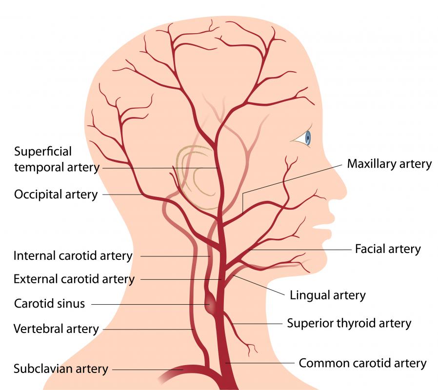
What are the Blood Vessels of the Brain? (with pictures) from images.wisegeek.com This vessel supplies blood to the front part of your brain, knows as your frontal lobe. Towards the anterior side of the brain, those arteries are the internal carotid arteries. Label the blood vessels in the inferior view of the brain using the hints provided. The carotid arteries and the vertebral arteries anterior cerebral artery (aca): Blood vessels are referred to collectively as the vascular system and, together with the heart, make up the circulatory system or cardiovascular system. Another whole article within the blood vessels and csf section is dedicated to the cavernous sinus. In the article on the ventricles within the cns, we will discuss their structure and. These vessels transport blood cells, nutrients, and oxygen to the tissues of the body.
Using medaka ( oryzias latipes ) as a model, the current protocol presents a quick and direct technique to label blood vessels in brain and pituitary by.
The structure, distribution and labeling of the whole brain vascular system of different arteries and veins in 3d. He says the restricted vessels prevent the blood from draining fast enough and injure the brain by causing a build up of iron which leads to ms. Label the blood vessels of the male pelvis using the hints provided. Over 1 year ago version: The blood vessels are the components of the circulatory system that transport blood throughout the human body. Label the veins of the anterior forearm and hand. In the article on the ventricles within the cns, we will discuss their structure and. Internal carotid artery (anterior circulation), vertebral artery (posterior circulation), and their hexagonal anastomotic network called blood brain barrier refers to the wall between the brain tissue and blood vessels. The difference in the structural characteristics of arteries, capillaries and veins is attributable to their respective functions. If you use this material, please attach a link to the artwork, i would really love to see it :d. Only some of the vessels that exist in a real brain have been labeled. The 500 ms patients, both adults and children, also underwent mri scans of the brain to measure iron deposits in surrounding areas of the brain. Fill in the blanks with the appropriate words to describe blood flow from the heart.
Comes off the subclavian a., ascends although the internal carotid a. Blood travels from the heart in arteries, which branch into smaller and smaller vessels, eventually becoming arterioles. The precise relation between blood vessels and brain regions, reflecting the physiology and pathology of brain function directly and accurately, has remained largely unknown. Blood vessels are critical to deliver oxygen and nutrients to all of the tissues and organs throughout the body. Internal carotid artery (anterior circulation), vertebral artery (posterior circulation), and their hexagonal anastomotic network called blood brain barrier refers to the wall between the brain tissue and blood vessels.
Science Source - Brain blood vessels, artwork from www.sciencesource.com Supplies the anterior brain and the vertebral a. The two cell types ensure the integrity of the neural vasculature by maintaining the blood. The blood vessel wall is endowed with connective tissue, smooth muscle, and striated muscles. The brain and its surrounding blood vessels exist in a close relationship. These vessels transport blood cells, nutrients, and oxygen to the tissues of the body. The carotid arteries and the vertebral arteries anterior cerebral artery (aca): Label the blood vessels in the inferior view of the brain using the hints provided. The precise relation between blood vessels and brain regions, reflecting the physiology and pathology of brain function directly and accurately, has remained largely unknown.
Function and homeostasis of the brain relies on communication between its complex network of cells.
The precise relation between blood vessels and brain regions, reflecting the physiology and pathology of brain function directly and accurately, has remained largely unknown. These vessels transport blood cells, nutrients, and oxygen to the tissues of the body. Fill in the blanks with the appropriate words to describe blood flow from the heart. Label the veins of the anterior forearm and hand. They also take waste and carbon dioxide away from the tissues. In the article on the ventricles within the cns, we will discuss their structure and. Blood in the brain is supplied by two pairs of large blood vessels (arteries): Blood vessel endothelium is continuous with the inner tissue lining of organs such as the brain, lungs, skin, and heart. About 2 years ago updated: This is particularly important structure due to its clinical implications, which are discussed in more detail in the article. If you use this material, please attach a link to the artwork, i would really love to see it :d. Label the blood vessels in the inferior view of the brain using the hints provided. The two cell types ensure the integrity of the neural vasculature by maintaining the blood.
Blood is also supplied to the brain by the vertebral a blood vessels labeled. This vessel supplies blood to the front part of your brain, knows as your frontal lobe.


0 Komentar